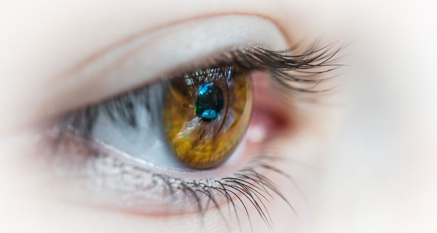
Understanding exactly how the eye works can be difficult. We know that the eye is responsible for vision, but how exactly is seeing possible? Even the experts of optometry had to spend years studying this and the basic anatomy of the eye. Unfortunately, not everyone has years to get the full picture, but this guide on basic eye anatomy can help.
The Basics of the Pupil
We all know what the pupil is, but do you know what the tiny black dot at the center of the eye does? The pupil is responsible for controlling the amount of light that enters the eye. This is an extremely important task, as everything we see has to do with light. When a pupil is larger than normal, this means that an individual takes in more light.
Just think about what happens after your pupils are dilated for an eye exam; the doctor hands you a pair of those stylish sunglasses in order to protect your eyes from light. Dilated pupils mean larger pupils, which means more light enters the eye. On the flip side, a smaller-than-average pupil takes in very little light. If you’re noticing anything strange going on with your pupils – like if they’re larger or smaller than normal or if they are two different sizes – have this checked out by your eye doctor.
The Basics of the Cornea
Another part of the eye that has to do with light consumption is the cornea. The cornea is located at the front of the eye and takes a lot of responsibility for the eye’s ability to focus light. It is composed of tissues that work to adjust the amount of water inside the cells located in the eye.
According to the Pacific University of Oregon, “the cornea is very sensitive to pain (think of how it feels when dust particles or a small eyelash get into your eye) because it has the highest density of nerves in the body. The cornea is also involved in keeping the surface of your eye moist with tears.” If you’re experience dry eyes, there’s a good chance you have a cornea irregularity. Most cornea-related eye issues can be treated fairly easily, especially dry eyes.
The Basics of the Refracting Tissues
Refracting tissues are responsible for refracting light as it enters the eye so that it is less harsh on nerve tissues that are sensitive to light (which are located at the back of the eye). Refracting tissues that function properly lead to clear, sharp, 20:20 vision. Problems with your eye’s refracting tissues can cause blurry vision.
The refracting tissues can be broken down into three categories: the pupil, the iris, and the lens. We’ve already talked about the pupil, which is located in the center of the iris (the colored portion of the eye). The iris is a muscle that helps to control pupil size. All of these eye structures and more play a role in vision.
If you found this article helpful, feel free to comment below or contact us at Tutor Circle!


Leave a Reply